Protect Your View
with a retina screening exam
What is retinal screening?
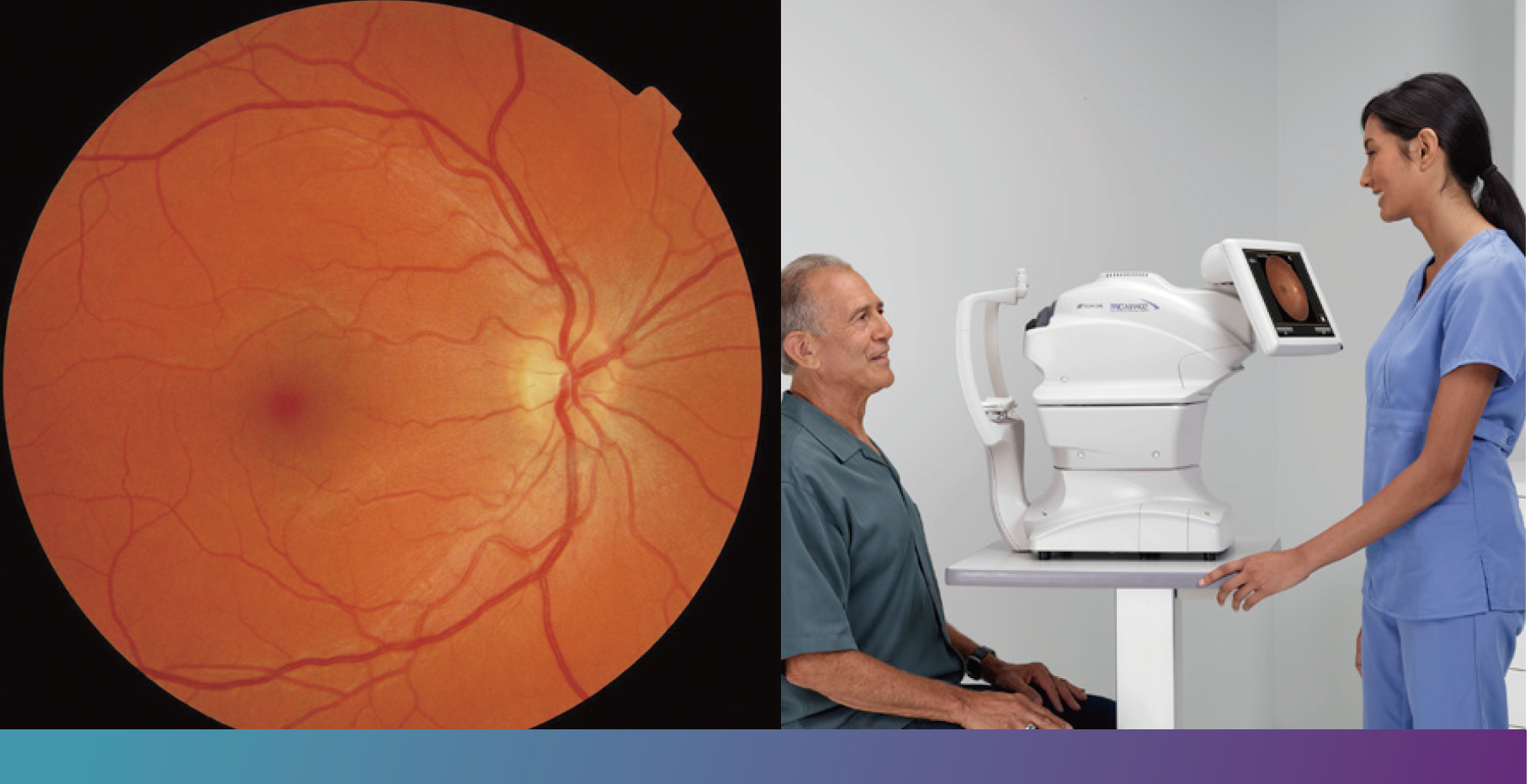
Retinal screening gives your doctor a high-resolution photograph of the retina, the light-sensing tissue at the back of your eye. The photograph shows detailed images of the structures of the retina to help identify early signs of disease before you begin to experience vision problems.


This new technology can greatly aid your doctor’s ability to accurately diagnose and document many diseases. It also provides a baseline for comparison with previous and future visits, which aids in monitoring disease progression and response to therapy.
Is a retina screening exam uncomfortable?
No, a retina photograph is a quick and painless process.
How long does an exam take?
A retina screening exam can be completed in just a few minutes.
Who should have a retinal screening?
Any person diagnosed with diabetes.
People with diabetes are at high risk for developing a variety of eye diseases including cataract, glaucoma, and diabetic retinopathy, one of the leading causes of blindness. Diabetic retinopathy is caused by damage to the small blood vessels in the patient’s retina. Early detection and treatment is a key to preventing vision loss, making it crucial for diabetic patients to have regular photographs taken of their eyes.

Patients may not have any symptoms in the early stages of the disease¹

95% of vision loss can be prevented with early detection²

Protect your vision with an annual comprehensive dilated eye exam
5 Advantages of Digital Retinal Screening

ONE
Your images are available immediately, allowing for quick diagnosis and an appropriate management for your unique case.
TWO
The images can be enhanced using sophisticated software to highlight any problem areas. This helps your doctor with diagnostic decisions.


THREE
Your doctor can show you your images at the time of your screening so you can make the most informed decision on how to manage your diabetes. Specialized software can help a family member understand in what way your vision may be affected and how you see your world.
FOUR
Digital photographs can be sent electronically to a co-managing doctor, if appropriate, allowing for more timely diagnosis & treatment.


FIVE
Digital photographs can be placed in your permanent clinical record, allowing your doctor to closely monitor even the slightest progression of any abnormalities.
Frequently Asked Questions
This exam consists of taking pictures of your eyes with a special camera to determine whether your diabetes is affecting your eye health. This is a medical screening, not a routine vision exam to determine whether you need vision correction. Patients with diabetes are required to have an annual diabetic retinal exam to prevent vision loss.
Many patients confuse routine eye exams with medical eye exams. During a routine eye exam, a doctor performs tests to determine whether a patient needs corrective lenses. A medical eye exam to screen for diabetic retinopathy uses different techniques to examine overall eye health. This diabetic retinal exam empowers your provider by providing a more comprehensive understanding of how your diabetes is affecting your whole body. Irregularities found in the eye are a key indicator to the overall effects of diabetes throughout the body.
Diabetic Retinopathy is the most common eye disease in diabetic patients and is the leading cause of blindness in adults in the United States. One in three people living with diabetes has some degree of diabetic retinopathy, and every person who has diabetes is at risk of developing it. There are no symptoms of diabetic retinopathy until it reaches an advanced stage. Diabetic Macular Edema is a complication of diabetic retinopathy and can also lead to additional vision loss and blindness. This screening will detect indicators related to both diseases, as well as other diseases or conditions of the eye that may require treatment.
Diabetic retinopathy develops when elevated blood sugar levels damage the blood vessels in the retina. Without detection and treatment, diabetic retinopathy leads to vision loss and eventual blindness.
Dilation opens the pupil to enable us to view the retina at the back of your eye. To capture quality retinal images, the technician will dilate your pupils naturally – without dilation eye drops. For most patients, sitting in a dark room for up to a few minutes with eyes closed is all that is necessary to sufficiently open the pupils for this screening.
There is no pain or discomfort experienced during this screening. You will experience a bright flash when the image is captured, similar to flash photography. The eye exam at your eye doctor that blows a puff of air is for a different test which checks for glaucoma.
Your exam images are interpreted by a board-certified eye care specialist. The results of the exam will be returned to your provider within a couple of days and your provider will be able to review the results with you.
If diabetic retinopathy is detected, your provider will determine next steps which may include a referral to an eye specialist for treatment.
Most insurance carriers allow this exam more than once every calendar year as part of your comprehensive diabetic care plan. Coverage of diabetic retinopathy screening varies by payer. We recommend that you check with your insurance provider to verify reimbursement eligibility prior to screening. The exams and billing codes used at an ophthalmologist’s office are different from the codes used at your primary provider’s office. Instances of duplicate charges are unlikely. However, if you have already had a full diabetic eye exam within this calendar year with an ophthalmologist, please provide us with the name of the doctor you saw so that we can obtain a copy of your exam report.
Insurance coverage will vary depending on your specific plan. Depending on your insurance policy, you may be responsible for paying a copay or deductible as with other services. Your insurance carrier will have more information about the coverage they offer.
